1. Introduction
N6-methyladenosine (m6A) is the predominant internal modification of eukaryotic messenger RNA, recognized as a dynamic and reversible mark that has transformed the understanding of post-transcriptional control. This mark is established by a nuclear writer complex centered on METTL3–METTL14 [1], removed by demethylases FTO and ALKBH5 [2,3], and interpreted by reader proteins that can influence the fate of methylated RNAs in a context-dependent manner. Early studies linked YTHDF2 to mRNA decay and YTHDF1 to translational enhancement [4,5], while the IGF2BP family is associated with transcript stabilization [6,7]. However, these roles should not be viewed as universally fixed; the degree of functional specialization, target overlap, and redundancy among cytoplasmic YTHDF paralogs remain a topic of ongoing debate [7–9]. Consequently, a single chemical tag can be related to splicing, export, stability, or translation outcomes, depending on the transcript, cellular state, reader context, and associated cofactors.
The context-dependent nature of m6A is particularly evident in cancer, where a specific regulator may function as an oncogene in one scenario while serving as a tumor suppressor in another. For instance, FTO promotes acute myeloid leukemia development by demethylating ASB2 and RARA, whereas ALKBH5 supports glioblastoma stem-like cell maintenance via FOXM1; however, both enzymes can inhibit tumor proliferation in other tissues. This dichotomy is often described as a "double-edged sword," accurately reflecting the contradictory findings without elucidating the factors that dictate the direction of the effect.
Two unresolved conflicts illustrate the limitations of this perspective, particularly within urinary system cancers. First, individual regulators can reverse their effects within the tumor cell compartment; for example, FTO stabilizes PGC-1α to suppress clear-cell renal cell carcinoma [2], while ALKBH5 stabilizes AURKB to promote the same disease [3]. Second, and less frequently acknowledged, m6A operates intrinsically within immune cells, where its net impact on antitumor immunity often contrasts with its effects in tumor cells. Loss of METTL3/METTL14 in tumors enhances CD8+ T cell infiltration and interferon-driven chemokines [10]. Deletion of YTHDF1 in dendritic cells improves antigen cross-presentation and enhances checkpoint blockade outcomes [11]. Conversely, tumor-intrinsic FTO may facilitate resistance to anti-PD-1 therapy [12]. This complexity suggests that a regulator promoting tumor cell growth may, in the surrounding immune compartment, either aid or inhibit the immune response against the same tumor.
Existing reviews often address related topics in isolation. Some characterize the tumor cell as a "double-edged sword," while others survey m6A's role in tumor immunity across multiple cancers or focus on a single writer or immune lineage. A comprehensive account that consolidates the mark's opposing net effects across tumor and immune compartments in a defined clinical context is lacking. Bladder, renal, and prostate cancers are particularly suitable for this purpose, as they encompass a broad range of immunogenicity, utilize checkpoint inhibitors, and have m6A-regulator signatures that track anti-PD-L1 response in urothelial carcinoma [13]. This grouping reflects a genuine translational thread rather than mere anatomical convenience. Renal cell carcinoma is among the most responsive to checkpoint therapies, while prostate cancer is among the least responsive, with urothelial carcinoma occupying an intermediate position and benefiting uniquely from intravesical BCG immunotherapy. A compartment-resolved account of m6A thus speaks to a tangible gradient of immune responsiveness within a single clinical specialty. However, this clinical gradient should not be misconstrued as evidence for a monotonic tumor-type-specific shift in the predominant m6A determinant. Available studies vary in model systems, molecular subtypes, immune-cell coverage, and endpoints, lacking a matched cross-cancer comparison that could rank the relative contributions of tumor-cell and immune-cell m6A programs. Consequently, it is inappropriate to infer that one determinant or one compartment becomes increasingly important from prostate to bladder to renal cancer. Instead, these three malignancies offer deliberately contrasting clinical and immunological contexts to test the same compartment-resolved framework. Their comparative value lies in assessing whether a model measuring tumor-cell and immune-cell m6A separately provides more insight than a bulk-tumor model across varied treatment environments, rather than presuming a fixed hierarchy of m6A mechanisms among cancer types.
This review reconciles the cell-autonomous and immune-cell-intrinsic roles of m6A in bladder, renal, and prostate cancer. It begins by summarizing how a single mark can yield opposing outputs and then contrasts evidence from tumor and immune cells across the three diseases. From this contrast, a framework for the "sign-flip" is proposed, taking into account the cell of origin, target transcript, reader availability, hypoxic and metabolic state, and spatial organization that collectively determine the direction of each effect. The discussion concludes by exploring how this duality influences responses to immunotherapy and resistance, and whether m6A-targeted agents such as METTL3 [14] and FTO inhibitors [15] can be effectively utilized alongside checkpoint blockade without incurring the compartment-level off-target liabilities predicted by the duality.
2. The m6A Toolkit and the Principle of Context-Dependent Output
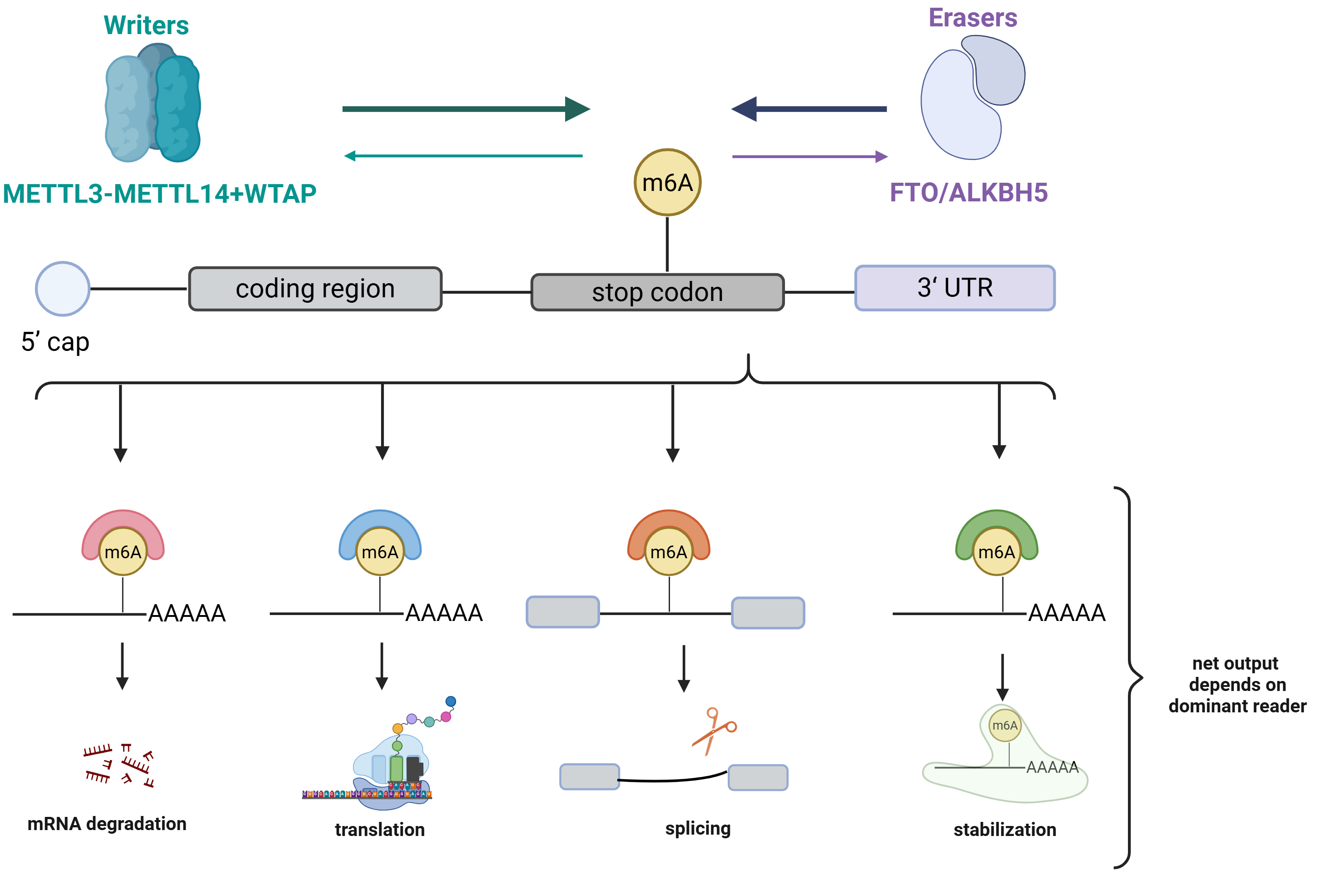
The regulatory logic of m6A is founded on three key activities: deposition, removal, and interpretation. Methylation is catalyzed by a writer complex centered around the METTL3–METTL14 heterodimer [1], while the removal of the modification is mediated by the demethylases FTO and ALKBH5 [16,17]. The m6A mark localizes near stop codons and in 3′ untranslated regions, labeling thousands of transcripts across the coding genome [18,19] (Figure 1). Importantly, the mark itself does not carry a fixed instruction; instead, its impact is assigned downstream based on reader engagement at specific sites under particular cellular conditions. This distinction between the presence of m6A and its interpretation forms the conceptual foundation for the compartment- and context-dependent behaviors discussed below.
The processes of writing and erasure render m6A a dynamic attribute of the transcriptome. The METTL3–METTL14 complex, along with its accessory subunits, deposits methyl groups co-transcriptionally [1], while FTO and ALKBH5 act to reverse this modification. The steady-state methylation level of any transcript thus reflects a balance between these opposing enzymatic activities [16,17]. This balance determines the fraction of a transcript pool that is modified at any given moment. Consequently, the same enzyme can either increase or decrease the abundance of a target, depending on the specific sites it modifies and the density of methylation. This stoichiometric characteristic indicates that neither writer nor eraser possesses an intrinsic direction of effect; each can be oncogenic or inhibitory based on the transcripts it regulates.
The reader layer links m6A to various post-transcriptional outcomes, but reader identity should not be equated with a fixed molecular fate. Early studies associated YTHDF2 with mRNA decay and YTHDF1 with translational enhancement [4,5], while YTHDC1 governs nuclear splicing and export [12], and IGF2BP proteins frequently stabilize their targets [6]. However, subsequent research using a unified YTHDF model reported significant overlap among YTHDF1, YTHDF2, and YTHDF3 targets, revealing a predominantly redundant, decay-promoting function in the examined cellular systems [7]. Further genetic studies have uncovered dosage- and cell-context-dependent compensation among these paralogs [8], alongside mechanistic analyses supporting distinct molecular and functional differences between YTHDF1 and YTHDF2 [9]. These competing observations suggest that a straightforward YTHDF1-translation/YTHDF2-decay dichotomy has not been established as a universal principle. Consequently, a reader–transcript mechanism should only be inferred when reader engagement, the resulting changes in target stability and/or translation, and the phenotype have been tested in the relevant cellular state. Factors such as reader abundance, localization, cofactors, transcript structure, m6A position and stoichiometry, as well as competition from other RNA-binding proteins, serve as potential contextual modifiers of reader occupancy and biological output [20]; these should not be interpreted as a fixed hierarchy of determinants. Additionally, potential reader-independent consequences of m6A must be considered when reader perturbation does not sufficiently explain observed molecular or phenotypic effects. Bulk m6A measurements average across these interacting variables, which clarifies their limited correlation with phenotype. Therefore, the term "reader context" is employed throughout this review, avoiding the assignment of fixed and mutually exclusive fates to individual YTHDF paralogs.
3. Tumor Cell–Intrinsic m6A Functions: Opposing Outcomes from the Same Regulators
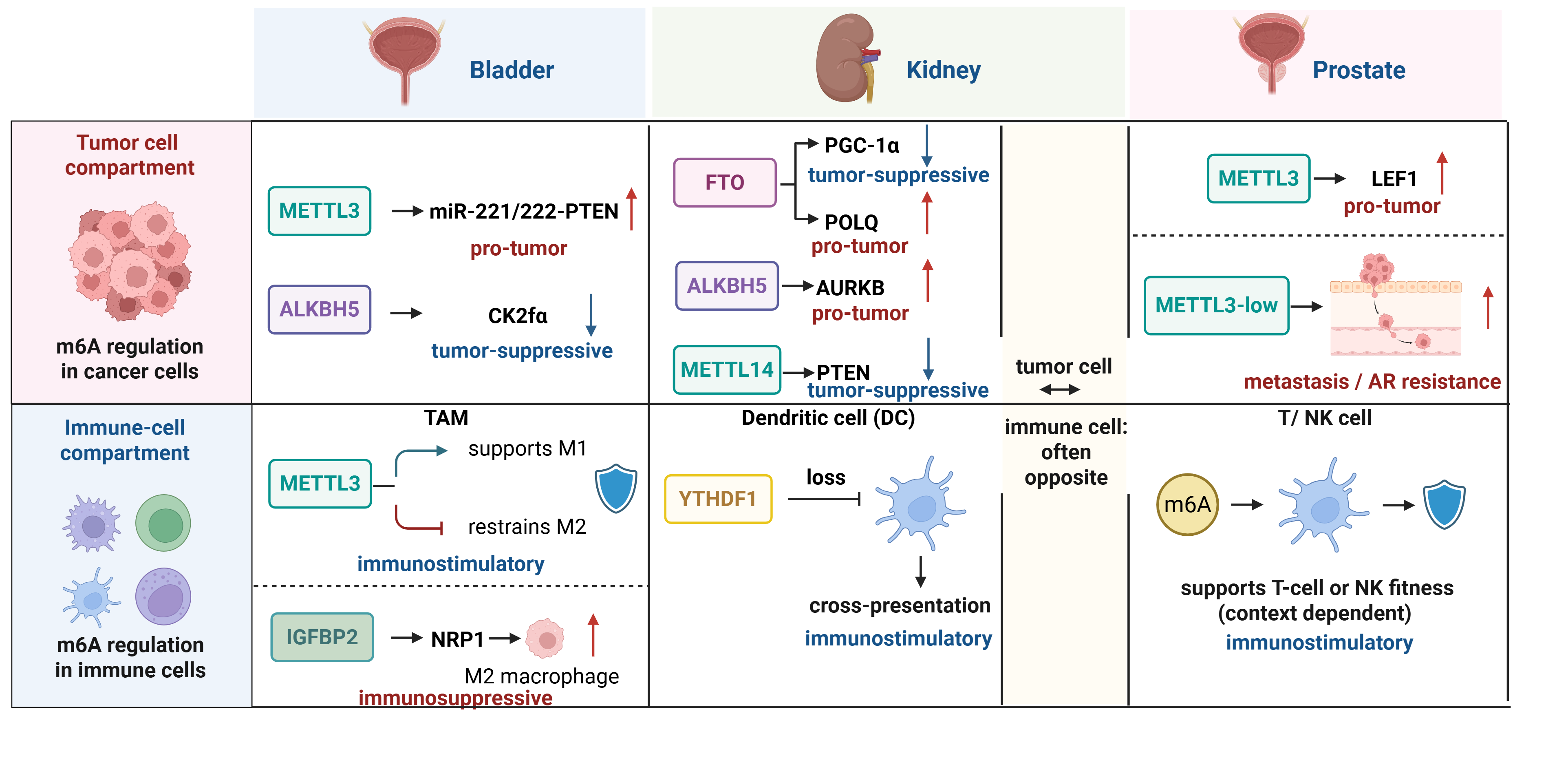
In tumor cells from bladder, renal, and prostate cancers, m6A regulation does not consistently exhibit a uniform direction of effect. In fact, an enzyme or reader may both promote and inhibit malignancy, depending on tumor subtype, the transcripts it regulates, and the repertoire of readers that interpret the associated marks. The strongest evidence emerges when examining a single regulator across multiple studies focusing on a specific disease, revealing opposing phenotypes that arise from distinct molecular targets. Cataloging these reversals within the tumor cell compartment establishes one facet of the duality explored in this review. Furthermore, it provides the mechanistic foundations upon which the subsequent framework explains the determinants of each effect's sign. The term "net effect" is utilized here to denote the direction of change in tumor growth and, where reported, in antitumor immune infiltration. Given that these outcomes can diverge, specific measured outcomes are indicated when pertinent. Evidence is evaluated in the context of the model system, employing causal language only in reference to in vivo conditional or functional perturbations, while regarding patient-cohort expression correlations as associations. The following description progresses from renal to bladder to prostate cancer, treating the opposing behaviors of each regulator not as a single canonical role but rather as a matched pair (Figure 2; Table 1).
3.1. Renal Cell Carcinoma
No regulator exemplifies directional flexibility as distinctly as FTO in renal cell carcinoma. Unlike PGC-1α, FTO acts as a tumor suppressor by demethylating and stabilizing the transcript, which restores oxidative metabolism and limits clear cell growth [2]. Conversely, when POLQ is targeted, FTO switches to an oncogenic role, enhancing proliferation and DNA damage tolerance [21]. In VHL-deficient tumors, which represent the majority of clear cell cases, FTO reveals a distinct function as a selective vulnerability, as cells depend on its demethylase activity [22]. Thus, its potential as a drug target is dictated by tumor genotype rather than a fixed classification as either an oncogene or a suppressor. METTL3 displays a similar conditional logic. Knockdown of METTL3 leads to increased cell proliferation, initially suggesting a tumor-suppressive role [23]. However, under sustained hypoxia, it engages in an HIF-1α–METTL3–PLOD2 axis that promotes tumor progression [24]. When analyzing the eraser and writer families, no consistent polarity is observed. For instance, ALKBH5, another eraser, facilitates renal carcinoma by stabilizing AURKB [3], while METTL14—like METTL3—has an opposing effect, maintaining PTEN to inhibit the PI3K–AKT pathway [25] and suppressing metastasis via Lnc-LSG1 [26]. Readers further complicate the regulatory landscape, predominantly favoring metabolic processes and tumor promotion. IGF2BP1 supports aerobic glycolysis by stabilizing LDHA [27], and a DMDRMR–IGF2BP3 complex promotes growth and metastasis through CDK4 and other cell cycle transcripts [28]. Decreased expression of YTHDF2 is linked to poor prognosis, emphasizing that even among readers, directional effects are not uniform [46]. Contradicting the notion that erasers are consistently oncogenic and writers reliably suppressive, data demonstrate otherwise. No single factor dictates direction; the impacted target transcript is central, while reader context and other cell-state variables can modify the outcome. Thus, neither class of writer, eraser, or reader, nor a fixed assignment of YTHDF paralogs, is sufficient to predict results.
3.2. Bladder Cancer
In bladder cancer, the balance shifts toward oncogenic signaling, while also providing examples that illustrate contrasting roles. The most influential actor is METTL3, which accelerates the conversion of pri-miR-221/222 to miR-221/222, subsequently reducing PTEN [29]. Together with YTHDF2, METTL3 degrades the tumor suppressors SETD7 and KLF4 [30]. Targeted removal of Mettl3 in bladder cancer stem cells leads to diminished tumor growth and blood vessel formation, positioning the writer at the forefront of a proliferative program [47]. Readers amplify this pathway, as YTHDF2 represses the innate immunity sensor DDX58/RIG-I in cancer cells [31]. YTHDF1 enhances resistance to cisplatin via an RPN2–PI3K/AKT pathway [32]. IGF2BP3 increases the half-life of both CDK6 [33] and HMGB1 [34], contributing to enhanced cell proliferation and survival. FTO further supports the oncogenic program by stabilizing STAT3 [35] and acting through a miR-576/CDK6 axis [36]. Conversely, ALKBH5 functions as a suppressor by demethylating CK2α to limit glycolysis and sensitize cells to cisplatin [37]. Deficiency of the nuclear reader YTHDC1, which binds RNA, leads to more invasive behavior [38]. Both instances highlight that, even within a writer-dominated disease, the polarity remains target-specific rather than fixed.
3.3. Prostate Cancer
Prostate cancer introduces a unique aspect where the same regulator can exhibit opposing effects relative to disease state compared to its influence on specific targets. For instance, METTL3 acts oncogenically by methylating LEF1, thereby activating Wnt signaling [39]. In contrast, low METTL3 expression is associated with advanced metastatic disease and confers resistance to androgen receptor antagonists [40], indicating that both high and low writer activity can correlate with aggressive disease depending on the context. Furthermore, the writer complex is regulated by androgens, thereby linking m6A deposition to the predominant oncogenic signals of the disease [48]. In this setting, FTO functions as a tumor suppressor. Its downregulation is indicative of higher tumor grade and increased invasiveness [41], and it stabilizes CLIC4 to mitigate growth [42]. The readers in this context favor tumor progression: YTHDF2 promotes degradation of the suppressors LHPP and NKX3-1, enhancing AKT signaling [43]. This effect is reciprocally constrained by miR-493-3p targeting YTHDF2 [49] and is further amplified by OTUB1, which stabilizes YTHDF2 protein50. Additionally, YTHDF1 increases the translation of PLK1, thereby activating the PI3K–AKT pathway [44]. The integration of these effects occurs through the androgen axis; under androgen deprivation, m6A-mediated repression of SIAH1 elevates CPSF1 levels, driving splicing toward the constitutively active AR-V7 variant and linking the m6A pathway to castration resistance [45].
Collectively, these tumor cell observations suggest a limited set of variables that determine the direction of effects: the cell of origin and molecular subtype, the transcript a regulator targets, the interpreting reader, and the tumor's metabolic or hypoxic state. The IGF2BP readers repeatedly appear in contexts favoring metabolic processes and cell-cycle stabilization, contrasting with decay-promoting readers that silence tumor suppressors. Consequently, reader composition often serves as the proximate determinant of whether a writer or eraser is perceived as oncogenic. Most supporting studies remain single-cohort correlations, with limited functional comparisons among competing targets. Only a few studies have investigated which effect prevails when a regulator affects both oncogenic and tumor-suppressor transcripts simultaneously, hindering accurate predictions about overall behavior based solely on expression levels. These factors primarily reflect processes within the cancer cell. Table 1 illustrates trends across the three cancer types. Erasers and writers cannot be categorized strictly by family; FTO, ALKBH5, METTL3, and METTL14 each appear on both sides of the table. The IGF2BP readers are predominantly found in the oncogenic column due to their roles in metabolism and cell-cycle stabilization, while decay-promoting readers more frequently silence tumor suppressors. Importantly, these same regulators also function within the immune cells surrounding tumors, where their net effects on antitumor immunity often differ. The comparison between these two compartments, rather than focusing on just one, is the primary objective of the next section and the supporting framework.
4. Immune Cell Intrinsic Roles of m6A in the Urologic Tumor Microenvironment
m6A regulators present in tumor cells are also found within the immune cells of the urologic tumor microenvironment, where their net effect on antitumor immunity often contrasts with their impact in the malignant compartment. This immune role is not merely a byproduct of tumor cell methylation; rather, it constitutes an independent layer of control that influences macrophage polarization, T-cell fitness, and dendritic-cell antigen presentation. Currently, checkpoint inhibitors are standard treatments for bladder and renal cancers, with ongoing studies in prostate cancer. Consequently, the nature of these immune-intrinsic effects directly impacts therapeutic responses. Direct, disease-specific mechanistic evidence is primarily concentrated in bladder cancer and remains limited in scope. The mechanisms involving macrophages, T cells, dendritic cells, and NK cells discussed below encompass both urological studies and cross-tumor mechanistic evidence; where cross-tumor data inform urological hypotheses, they are identified as extrapolative rather than established mechanisms in bladder, renal, or prostate cancer (Figure 2; Table 2).
4.1. Tumor-Associated Macrophages
Most macrophage-intrinsic mechanisms mentioned were elucidated in non-urological experimental systems and should be interpreted within a cross-tumor mechanistic context. The strongest direct evidence in urology pertains to bladder cancer, summarized separately below. In tumor-associated macrophages, the writer METTL3 typically counteracts the immunosuppressive M2 program that promotes tumor growth. The m6A deposited by METTL3 restrains the translation of Snail, suppressing M2 polarization, while macrophage-specific loss of METTL3 accelerates tumor growth [52]. In parallel, METTL3 methylates STAT1 to enhance the pro-inflammatory M1 state [53]. The balance between M1 and M2 significantly shapes the immunosuppressive environment within urologic tumors; thus, a writer favoring M1 macrophages serves as an intrinsic restraint on tumor-promoting inflammation. The removal of this writer from the myeloid compartment highlights this relationship. Deletion of myeloid Mettl3 impairs YTHDF1-dependent translation of SPRED2, hyperactivates NF-κB and STAT3, expands regulatory T cells (Tregs), and diminishes the response to anti-PD-1 therapy [54]. In this context, the writer acts as a brake on pro-tumoral myeloid reprogramming, contrasting with its typically oncogenic role within tumor cells, exemplifying the compartmental opposition documented in this section.
Evidence for reader and eraser effects in macrophages exhibits significant heterogeneity. Currently, direct disease-specific evidence is largely restricted to bladder cancer, while several well-cited macrophage mechanisms originate from non-urological tumor models. In bladder cancer, the reader IGF2BP2 stabilizes NRP1, driving M2 polarization and malignant behavior, which positions the IGF2BP2–NRP1 axis as a therapeutic target specific to this compartment [51]. In cross-tumor models, loss of the reader YTHDF2 reprograms macrophages toward an antitumoral phenotype through IFN-γ–STAT1 signaling, enhancing CD8+ T cell priming [55]. In non-urological models, the demethylase ALKBH5 fosters M2-like, immunosuppressive states within the tumor microenvironment [56]. For example, in non-small cell lung cancer, ALKBH5 modulates tumor–macrophage interactions to recruit PD-L1-expressing M2 macrophages, thereby affecting susceptibility to anti-PD-L1 therapy [57]; however, its relevance to renal and urothelial tumors has yet to be directly tested. The influence of m6A extends to non-coding intermediates. In colorectal cancer models, m6A modification of miR-146b reshapes tumor-associated macrophages and enhances anti-PD-1 efficacy, demonstrating that this modification can reverse an immunosuppressive myeloid program [58]. A bladder-specific study further elucidates the relationship between tumor and immune compartments. METTL3 contributes to an immunosuppressive microenvironment by upregulating CXCL5 and suppressing CCL5, thus recruiting myeloid-derived suppressor cells (MDSCs) and reducing CD8+ T cell infiltration, which restores immune balance and enhances the anti-PD-1 response [50]. A single writer can exhibit pro-tumoral effects in bladder cancer cells while also influencing the surrounding immune context through myeloid and chemokine interactions. Therefore, the compartment must be specified prior to determining directionality. The bladder METTL3 studies provide the strongest urologic evidence that the immune contribution can dominate. In the same context where METTL3 promotes tumor-cell proliferation, its inhibition enhances the anti-PD-1 response in conjunction with CXCL5/CCL5-dependent myeloid and chemokine remodeling [50]. This finding suggests that the therapeutic consequences of tumor-cell METTL3 perturbation can be mediated via the immune microenvironment. However, it does not quantify the relative contributions of tumor-cell-intrinsic versus immune-cell-intrinsic METTL3 activity. Thus, the bladder data offer a compelling rationale for compartment-resolved comparisons, rather than conclusively proving that the immune compartment invariably dictates the net effect of METTL3 blockade.
4.2. T lymphocytes and Regulatory T Cells
Except for tumor-cell-derived chemokine evidence in bladder cancer, the T-cell and regulatory-T-cell mechanisms summarized below were predominantly established in non-urological systems and should not be considered direct urological immune-cell evidence. T cells illustrate a stark contrast: while m6A is essential for their proper functioning, the absence of m6A in tumor cells enhances antitumor immunity. In conventional T cells, METTL3-dependent m6A modification regulates SOCS family transcripts, facilitating IL-7/STAT5-induced effector differentiation. In Tregs, m6A maintains the suppressive program via IL-2/STAT5 signaling [59,60]. Hence, m6A supports both aspects of the T-cell response depending on lineage. However, therapeutic strategies should focus on targeting the tumor rather than altering T-cell m6A. The inactivation of METTL3 or METTL14 in tumor cells stabilizes Stat1 and Irf1 signaling, leading to increased CD8+ T-cell infiltration and the recruitment of chemokines such as IFN-γ, CXCL9, and CXCL1015. The dynamics of this equilibrium become more nuanced when considering readers and erasers. YTHDF2, abundantly present in early effector CD8+ T cells, is crucial for controlling their polyfunctionality; its deletion promotes tumor progression and confers resistance to anti-PD-1 therapy [61]. ALKBH5 influences CD4+ T cell pathogenicity by regulating the stability of Ifng and Cxcl2 transcripts [62]. Furthermore, macrophage m6A also impacts the same circuit; loss of METTL14 in tumor-associated macrophages leads to CD8+ T-cell dysfunction due to the accumulation of Ebi3 [63]. The commonality across these lineages is that the type of cell in which m6A is perturbed significantly influences its immunological direction—whether pro- or antitumor—alongside the target transcripts, reader context, and local microenvironment. This interlinking of myeloid and lymphoid programs within the same microenvironment demonstrates that perturbations confined to one immune cell type can affect another.
4.3. Dendritic Cells and Natural Killer Cells
Dendritic cells and natural killer cells further complete the immune-intrinsic landscape and provide clear therapeutic proof of concept. Deletion of the reader YTHDF1 in classical dendritic cells reduces m6A-dependent translation of lysosomal cathepsins, enhancing cross-presentation of tumor antigens and CD8+ T-cell cross-priming, effectively synergizing with PD-L1 blockade16. Conversely, METTL3-mediated methylation facilitates dendritic cell activation by promoting translation of CD40, CD80, and the TLR4 adaptor Tirap [64]. This scenario highlights that a writer and a specific reader can operate in opposing directions within the same lineage. The dendritic cell axis thus provides significant insight, indicating that m6A governs the initiation of antitumor immunity and that different components of this machinery can be targeted for opposing effects. In natural killer cells, METTL3 sustains antitumor function through m6A-marked SHP-2 and IL-15–driven signaling; its absence diminishes NK cell infiltration and activity, accelerating tumor growth [65]. Across these lineages, the consistent pattern reveals that a regulator does not possess a singular immune role; its output is contingent upon the cell type and the transcripts being regulated, often signaling in the opposite direction compared to tumor compartments.
The immune compartment in prostate cancer represents a unique case and the weakest link in the urologic evidence, serving as a critical test of the framework's applicability. Prostate cancer is characterized as an immunologically cold tumor, featuring a low mutational burden, minimal tumor-infiltrating lymphocytes, and a microenvironment dominated by MDSCs and Tregs. This positioning places it at the low-responsive end of the immunogenicity gradient discussed in this review. So far, almost no studies have delineated cell-intrinsic m6A programs in prostate immune cells. The androgen axis is a pertinent avenue for exploration, as androgen signaling affects the functions of T-cell and myeloid cells, and the m6A writer complex itself is regulated by androgens [48]. Hence, it is hypothesized that androgen deprivation may reshape the m6A biology of prostate immune cells, paralleling its effects on tumor cells. This hypothesis represents a specific, testable prediction rather than an established mechanism, and all claims regarding intrinsic prostate immune function are labeled as extrapolations due to the absence of organ-specific data.
Collectively, the immune-intrinsic evidence constitutes the second half of the duality described throughout this review. Regulators such as METTL3, ALKBH5, and specific YTHDF-associated pathways exhibit opposing net effects in immune cells compared to tumor cells. Consequently, the overall impact of any m6A disruption in a urologic tumor emerges from the interplay of both compartments, which may either negate or amplify each other. A writer might inhibit M2 polarization and enhance dendritic cell priming while simultaneously promoting proliferation in adjacent cancer cells; thus, its significance as a drug target cannot be comprehensively assessed from either compartment alone. The implications for translational research are profound, as several of these pathways intersect with the efficacy of checkpoint blockade, and data from bladder chemokines and macrophages suggest that immune-directed effects may sometimes outweigh cell-autonomous effects. Nevertheless, the evidence base remains inconsistent and unevenly distributed across the three diseases. Direct urologic data predominantly focus on bladder cancer. Table 2 differentiates evidence derived from human urological specimens from mechanistic data that remains at the preclinical stage. Currently, human data largely consist of associations related to expression, immune infiltrate, or treatment response; direct demonstration of a cell-intrinsic m6A mechanism in sorted human immune cells is not yet available for bladder, renal, or prostate cancer. The renal immune microenvironment is primarily explored through tumor cell and pan-cancer studies, while the prostate immune compartment relies heavily on inferences rather than direct validation; therefore, claims regarding immune-intrinsic mechanisms in prostate cancer should be considered extrapolative until organ-specific evidence emerges. Few studies employ cell-type-specific deletions with the spatial resolution necessary to confirm the action of a regulator within an intact tumor. The next section addresses the challenges of reconciling these opposing compartmental effects into a disease-specific conceptual framework and identifying the determinants of the directionality of m6A perturbations in specific cells and diseases.
5. Reconciling Two Compartments: A Conceptual Model for the Sign-Flip
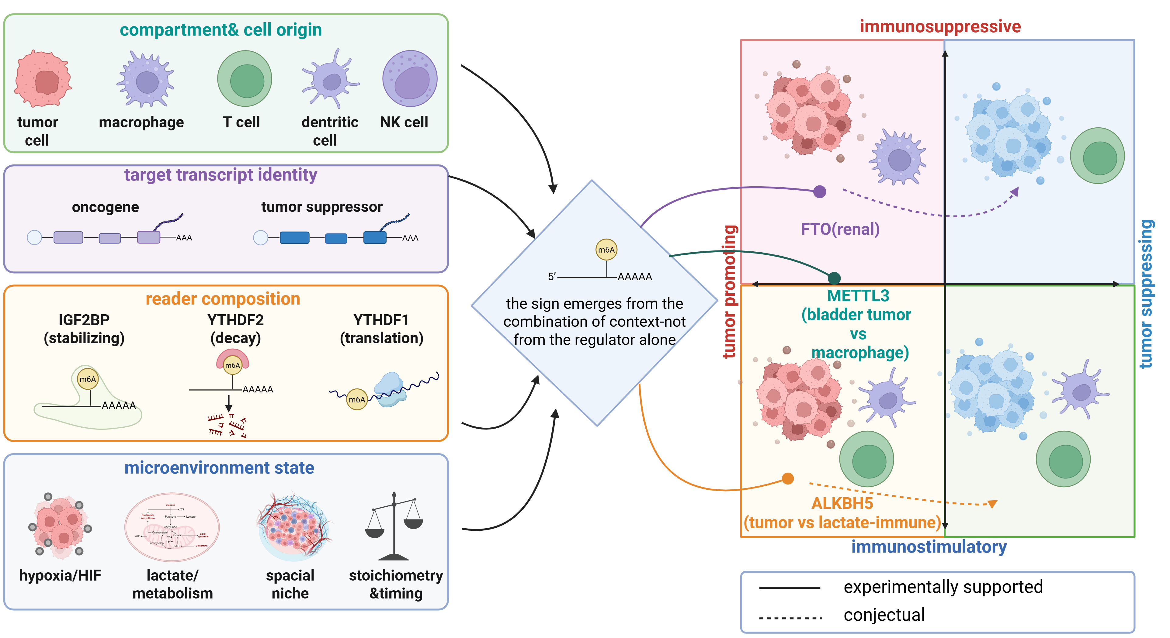
The opposing behaviors observed in tumor and immune compartments cannot be solely attributed to a regulator. Thus, a compartment-resolved conceptual model is utilized to organize the contextual variables that may influence the direction of an m6A perturbation (Figure 3). This model does not establish these variables as a validated decision rule or allow for prospective phenotype inference. Instead, it emphasizes a set of experimentally testable explanations: the cell type in which a regulator acts, the transcripts available in that cell, the readers interpreting modified RNA, and the metabolic and spatial state of the tissue. The model's specific aim is to clarify the potential opposition between tumor-cell and immune-cell effects, facilitating the separate measurement and perturbation of these components. To differentiate direct from indirect influences, the determinants within this framework are classified into proximal and contextual levels. Proximal determinants operate directly on m6A output at the RNA level, encompassing the identity of the modified transcript, the presence and stoichiometry of m6A at relevant sites, and the reader involved in determining the fate of the RNA—whether it is degraded, stabilized, translated, spliced, or exported. Contextual determinants, which include cell lineage, molecular subtype, hypoxia, metabolism, and tissue architecture, predominantly exert indirect effects. These factors reshape the expressed transcript pool, influence the recruitment or activity of writers and erasers, affect reader abundance or localization, and determine the signaling environment that shapes the interpretation of a proximal m6A event. Target-transcript identity thus occupies a critical position at the interface of these two levels: it serves as a direct determinant of phenotype once modified, while its availability and methylation status can be influenced by cell state and chromatin programs. For instance, H3K36me3 can recruit the METTL3–METTL14 complex to actively transcribed nascent RNA, guiding m6A deposition co-transcriptionally [66].
The compartment and cell of origin where a regulator functions serve as the primary axis for understanding various regulators, as a single enzyme can participate in different transcriptional programs across tumor cells and immune cell lineages. METTL3 exemplifies this duality. Initially, it promotes proliferation and an immunosuppressive chemokine program in bladder cancer cells [50], while subsequently restraining the pro-tumoral M2 state in macrophages [52]. Consequently, its overall impact on tumors is shaped by contributions from each compartment. Tumor subtype and disease stage represent finer gradations along this axis. For instance, METTL3 is oncogenic in localized prostate cancer but assumes a low-activity state in metastatic disease and antiandrogen resistance, indicating that even within a single malignant lineage, the differentiation state of the cell can alter effects [40]. Compartment, therefore, serves as the initial stratification for hypothesis generation, focusing not on what m6A does to cancer but on what a regulator does within a specific cell context.
The third and fourth factors, target transcript identity and reader context, are interrelated yet distinct. The functional outcome of altering a writer or eraser as oncogenic or suppressive primarily hinges on the biological role of the affected transcript. For example, FTO can stabilize the tumor-suppressive PGC-1α transcript in renal carcinoma while simultaneously supporting oncogenic pathways through other targets [35,67]. Reader context may influence the fate of a methylated target; however, current evidence does not support assigning a universal translational function to YTHDF1 or a universal decay function to YTHDF2. Some systems indicate paralog specialization, while others suggest significant overlap and functional compensation among YTHDF proteins [7–9]. Consequently, reader composition should be treated as a hypothesis-generating contextual variable that necessitates direct testing in the relevant cell state, in conjunction with reader localization, cofactors, target binding, RNA stability, and translation. The IGF2BP2–NRP1 axis in bladder-associated macrophages serves as a defined example, wherein a specific reader-target interaction facilitates M2 polarization [51]. Thus, measuring the abundance of writers or erasers alone rarely predicts phenotype, and a fixed assignment of YTHDF paralog functions cannot substitute for target-specific mechanistic evidence.
Microenvironmental state encompasses the final determinants, including metabolic and hypoxic conditions, spatial organization, and the stoichiometry and timing of methylation. Hypoxia can dramatically alter a regulator's function, sometimes resulting in opposing effects. For instance, within the context of the HIF-1α–METTL3–PLOD2 axis, METTL3 acts as a driver of progression in renal carcinoma under persistent low-oxygen conditions [24], contrasting with its suppressive role in normoxic environments. Metabolism serves as the link between the two compartments, as changes in eraser activity influence tumor lactate levels, thereby affecting the accumulation of suppressive myeloid and regulatory cells that determine immunotherapy outcomes [68]. This interplay illustrates cross-compartment integration, where a tumor-cell m6A perturbation can indirectly reshape adjacent immune-cell states through shared metabolic mediators. A modification in tumor cell m6A affecting a metabolic transcript can influence the extracellular lactate pool via transporters like MCT4, reprogramming nearby myeloid and regulatory cells without altering their own m6A status. Consequently, the methylation state of one compartment can exert an influence through metabolic pathways on another, emphasizing that evaluations of metabolic axis perturbations must consider more than just the tumor cell. Additionally, spatial organization introduces another dimension often overlooked by bulk analyses. The expression of regulators and readers varies across tumor core, invasive margins, and intratumoral immune aggregates. An enzyme may exhibit oncogenic properties when tumor cells predominate, while potentially acting in an immunostimulatory manner just a few hundred microns away in areas where macrophages cluster. This divergence is highlighted by the opposing consequences of METTL3 loss in tumor cells compared to its deletion in the myeloid compartment within the same microenvironment [54]. Timing and stoichiometry further influence these dynamics. Both the methylated fraction of a transcript pool and the specific activation or differentiation stage during which methylation occurs determine the functional relevance of a mark. For instance, FTO suppresses one renal target while promoting another within the same disease context [21], illustrating that these determinants operate in concert rather than allowing any single variable to dominate.
When compiled into a decision logic, these determinants form a structured basis for formulating and testing hypotheses regarding the sign of an m6A perturbation, enabling predictions that can be inferred rather than assessed only retrospectively. The process begins by identifying the compartment and cell type, followed by evaluating whether the engaged transcript is oncogenic or suppressive. The dominant reader interpreting the transcript is then identified, and the prediction is conditioned on the tissue's metabolic, spatial, and temporal state (Figure 3).
Three illustrative examples demonstrate the application of this logic. Observations suggest that FTO may suppress clear cell renal carcinoma by acting on PGC-1α in oxidative tumor cells, while simultaneously promoting growth through engagement with proliferative or DNA repair transcripts, which aligns with divergent findings in renal contexts. METTL3 is predicted to have a net pro-tumoral effect when its primary activity is in bladder cancer cells, yet a net immunostimulatory effect when its dominant role occurs in macrophages or dendritic cells. Thus, the therapeutic value of inhibiting METTL3 is contingent upon which compartment exerts the more significant influence. ALKBH5 presents another case highlighting the need for careful specification of pathway direction. In renal tumor cells, ALKBH5 promotes proliferation by regulating AURKB expression [3]. In contrast, studies in B16 melanoma and CT26 colorectal tumor models undergoing anti–PD-1 immunotherapy show that ALKBH5 loss or pharmacological inhibition increases m6A on Mct4/Slc16a3, decreases MCT4 expression and intratumoral lactate levels, and correlates with reduced Treg and MDSC accumulation, as well as improved treatment response [68]. These findings do not confirm that systemic ALKBH5 inhibition will yield the same immune effects in renal or other urological cancers; instead, they provide useful cross-tumor mechanistic context and require validation in urological models before inferring a combined benefit for both tumor and immune contexts. This framework challenges the "double-edged sword" metaphor, reframing it not as a lingering paradox but as a function with specified input conditions. The logic underpinning this concept is illustrated in Figure 3 as a conceptual map that progresses from the compartment and cell of origin to the microenvironmental state, with the three examples outlining distinct paths leading to their respective outcomes. The figure also delineates areas requiring further investigation, as solid and dashed lines differentiate steps supported by direct experimentation from those that remain inferred.
5.1. Predictions and Disconfirming Experiments
Currently, this framework serves as a conceptual and hypothesis-generating model rather than a validated quantitative predictor of therapeutic response or m6A function. Its practical value lies in organizing heterogeneous observations, pinpointing variables likely to clarify apparently conflicting results, and prioritizing experiments where the relevant compartment, target transcript, reader context, and microenvironmental conditions can be tested together. Direct perturbation experiments conducted under well-defined biological conditions remain the standard for establishing functional directionality. Integrating the four dimensions can enhance experimental design and interpretation, yet it cannot replace experimental evidence or reliably predict the net effect of any m6A intervention in an untested tumor context.
As a model rather than a narrative, this framework proposes falsifiable hypotheses that specifically designed experiments could potentially disprove. For instance, in one genetically modified mouse model, the deletion of METTL3 in epithelial and myeloid cells is expected to produce opposing effects on the density of intra-tumoral CD8+ T cells: the loss in epithelial cells would presumably increase this density through the induction of interferons and chemokines, while the loss in myeloid cells would decrease it due to M2 expansion. If both perturbations yield the same directional effect, the proposed compartment-specific interaction would not be supported in that model.
To assess the respective contributions of tumor and immune compartments, perturbations should be compared in matched immunocompetent orthotopic models. A tumor-cell-restricted arm would selectively perturb METTL3 in implanted cancer cells while preserving a wild-type host, whereas reciprocal immune-lineage-restricted arms would maintain METTL3-intact tumor cells in hosts with conditional deletions in myeloid cells, dendritic cells, or T-cell subsets. A combined-perturbation arm would determine whether the effects are additive, antagonistic, or non-linear. In each arm, target engagement should be quantified separately in sorted tumor and immune populations alongside measurements of tumor growth, anti-PD-1 response, chemokine output, macrophage states, CD8+ T-cell infiltration, and survival outcomes. Comparing the effect sizes across these conditions would help differentiate tumor-cell-autonomous effects from immune-cell-intrinsic effects, and reveal whether the observed therapeutic benefit originates from tumor-to-immune signaling.
Additionally, the proposed influence of reader context should be treated as an empirically testable hypothesis rather than an established mechanism. The relative abundance of IGF2BP and YTHDF proteins does not establish occupancy or biological output at a shared target, as these outcomes may depend on transcript structure, m6A position and stoichiometry, subcellular localization, cofactors, and competing RNA-binding proteins. A rigorous test should measure reader occupancy at defined methylated sites and compare outcomes from single-reader versus combined-reader perturbations. Subsequently, assessing target stability, translation, and phenotypic changes, along with conducting rescue experiments in the relevant cell state, would provide insights. Observing a phenotype reversal under these conditions would support a reader-context-dependent mechanism in that model; conversely, a lack of reversal would limit the applicability of the model without establishing a universal rule. Changes in tumor lactate or oxygen levels may also influence responses to eraser perturbations, yet this remains a conditional hypothesis requiring direct measurements of metabolic states, m6A alterations, and downstream effectors. Conditional, lineage-specific perturbation remains essential for delineating tumor-cell and immune-cell contributions in vivo. Thus, the framework does not yield a quantitative model of reader occupancy or compartment weighting; rather, it identifies experimentally falsifiable questions that can guide studies focused on elucidating mechanisms.
5.2. Measuring the Framework: Single-Base, Single-Cell, and Spatial Detection of m6A
The model prompts a measurement agenda, making its testability dependent on methods capable of determining m6A at specific sites. Although antibody-based mapping initiated the field, it also imposes limitations. Techniques like m6A-seq and MeRIP-seq report peak-level enrichment rather than pinpointing single-base positions, require bulk input, and cannot assign a methylation mark to a specific cell [18,19]. Two advancements now address the stoichiometric variable emphasized by the framework. GLORI employs glyoxal- and nitrite-mediated deamination of unmethylated adenosines to quantify m6A with single-base resolution, similar to how bisulfite sequencing quantifies DNA methylation [69]. m6A-SAC-seq generates comparable single-base quantitative maps through enzyme-assisted labeling of low-input RNA [70]. These methodologies make the fraction methylated term of the framework directly measurable rather than assumed.
Cell type resolution presents a more challenging requirement, yet antibody-free chemistry is beginning to meet this need. DART-seq integrates the cytidine deaminase APOBEC1 with an m6A-binding YTH domain to edit cytidines adjacent to methylated sites, enabling the reporting of m6A from as little as a nanogram of input [71]. By extending this design to single cells, scDART-seq maps m6A across thousands of individual cells, revealing high heterogeneity in methylation, often with many sites present in only a small fraction of cells [72]. This heterogeneity aligns with the model's premise and argues against inferring per-cell states from bulk data. Spatial detection, however, remains an underdeveloped frontier; currently, no technique exists for co-resolving reader composition and per-cell methylation in situ throughout the tumor core, invasive margin, and immune aggregates. Consequently, spatial predictions made by the framework cannot yet be targeted at scale. Addressing this gap represents the most significant methodological challenge that could confirm or disprove the sign-flip framework in intact urologic tumors.
These approaches should be viewed as a series of steps rather than a single assay. Single-base resolution quantification determines the stoichiometry term, single-cell mapping connects each mark to a specific compartment, and spatial profiling seeks to locate that compartment within the tissue. A logical strategy would thus involve pairing single-cell m6A mapping with matched reader and lineage markers in bladder, renal, and prostate tumors, subsequently introducing spatial context as the technology advances. This conceptual model can only be examined when the same tumor is analyzed at single-base, single-cell, and spatial resolution, allowing for the measurement of compartment-specific effects, spatial relationships, and intercellular interactions in relation to the observed outcomes.
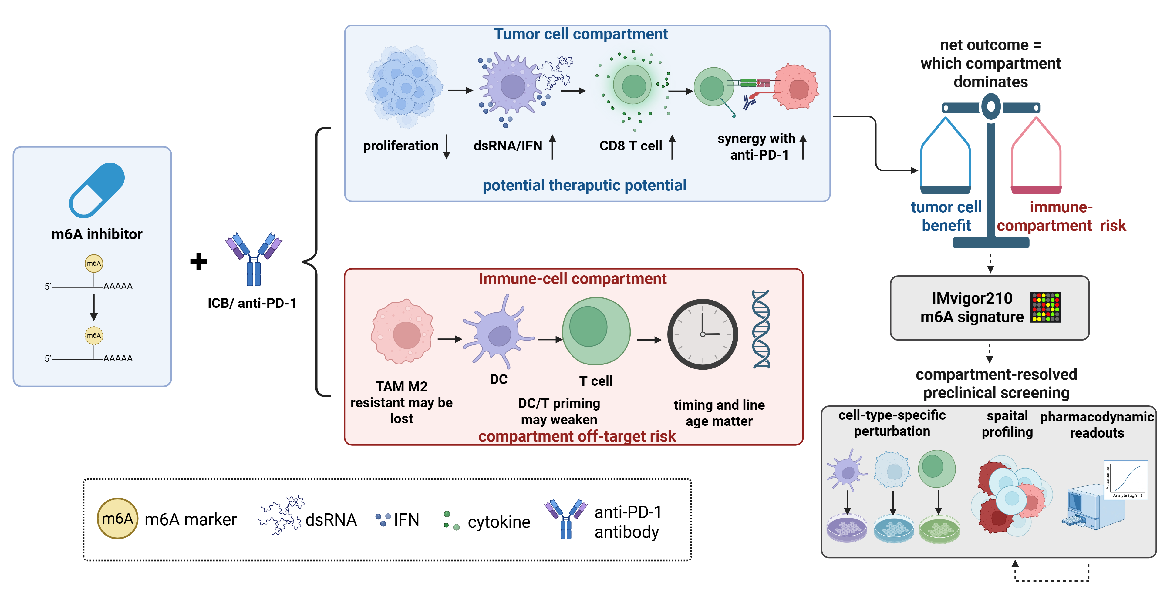
The compartment-resolved model promotes a translational hypothesis: systemic targeting of an m6A regulator may yield opposing effects in tumor and immune compartments. Since the net effect of an m6A regulator arises from the integration of contributions from both tumor cells and immune cells, including their interactions across space and time, the utility of m6A as a biomarker for immunotherapy response and the safety of m6A-targeted therapies depend on which compartment dominates in a given patient. Checkpoint blockade has become central to managing urothelial and renal carcinoma, is currently under investigation in prostate cancer, and preclinical studies indicate that m6A regulators influence responses to this therapy. However, the same duality that renders these regulators promising targets carries implications for preclinical design. An inhibitor that silences an oncogenic program in tumor cells might also inhibit a beneficial immune program, necessitating a screening of combination strategies at the compartment level rather than assuming their safety or efficacy based solely on tumor cell data (Table 3).
As biomarkers, m6A regulators currently track immunotherapy responses in the urologic context pertinent to this review. An m6A-regulator-based nomogram derived from the IMvigor210 atezolizumab cohort predicts anti-PD-L1 responses in advanced urothelial carcinoma, providing direct evidence that the m6A pathway is informative in bladder cancer [13]. Mechanistic studies elucidate why such markers hold significance. In bladder cancer, a methionine-driven YTHDF1 axis degrades RIG-I and enhances PD-L1 translation, thereby linking m6A reading to immune evasion and checkpoint ligand expression [73]. The clinical relevance of m6A regulators extends beyond bladder cancer. Renal cell carcinoma is among the most responsive solid tumors to checkpoint inhibition, whereas prostate cancer ranks among the least responsive, suggesting a natural gradient across which a compartment-resolved biomarker could be tested. However, it remains uncertain whether mechanisms identified in pan-cancer models apply to renal or prostate cancer, necessitating direct disease-specific validation. Tumor-intrinsic FTO, for instance, promotes resistance to anti-PD-1 therapy by regulating m6A levels on PD-1, CXCR4, and SOX10 [12], while ALKBH5 status influences responsiveness through its effects on tumor lactate and subsequent establishment of suppressive myeloid and regulatory populations [68]. Through the lens of the framework, these markers are informative specifically because they reflect compartment-specific states rather than a singular global level of methylation. This highlights the prediction that a signature trained in one disease might require recalibration before applicability to another context. More precisely, a valuable urologic biomarker is expected to consist of a deconvolved, cell-type-resolved measurement, wherein contributions from tumor cells and immune cells are assessed separately and then weighted according to the disease. The single-base and single-cell methods outlined previously are essential for making such biomarkers measurable, with the immunogenicity gradient spanning bladder, renal, and prostate cancer serving as a validation ground for predicted shifts in compartment weighting.
Pharmacological strategies have also advanced, with several agents now linking m6A inhibition to enhanced antitumor immunity. The catalytic inhibition of METTL3 by STM2457 was the first demonstration indicating that inhibiting m6A writers is a viable anticancer approach [14]. Subsequent studies revealed that enzymatic inhibition of METTL3 triggers a cell-intrinsic double-stranded RNA and interferon response that enhances T-cell-mediated killing and synergizes with anti-PD-1 [74,75]. Independent evidence supports the notion that targeting METTL3 potentiates T-cell antitumor functions [76]. FTO inhibitors have followed a similar trajectory, moving from inducing cell-intrinsic cytotoxicity to promoting immune activation. FB23-2 validated FTO as a druggable target in leukemia [77], while more potent inhibitors like CS1 and CS2 sensitized tumors to T-cell killing by decreasing the expression of the immune checkpoint LILRB4 [78]. Furthermore, Dac51 alleviated an FTO-driven glycolytic program, increasing the infiltration of CD8+ T cells and enhancing synergy with checkpoint blockade [15]. The evolution of the FTO series from a primarily cytotoxic approach to one encompassing metabolism and immunity resonates with the framework's assertion that the effects of a regulator in tumor cells and immune cells represent distinct targets. Reader-directed strategies are expanding this therapeutic arsenal; for example, targeting YTHDF1 reduces MDSCs and enhances the effectiveness of anti-PD-1 therapy [79]. However, most of these findings originate from leukemia, colorectal cancer, or melanoma models rather than urological tumors, indicating that they should be considered cross-tumor pharmacological rationale rather than definitive evidence of efficacy in bladder, renal, or prostate cancer.
The framework warns that systemic inhibition of a regulator targets all compartments simultaneously; thus, the therapeutic effect of a drug cannot be concluded from its impact on tumor cells alone. METTL3 inhibition exemplifies this conflict, as the same treatment that inhibits bladder cancer cell proliferation and disrupts a chemokine-mediated immunosuppressive program [50] also eliminates a writer in the myeloid compartment that governs M2 polarization and enhances dendritic cell priming; the loss of this function may promote tumor growth and decrease the anti-PD-1 response [54]. The net outcome's favorability toward the tumor or the patient depends on which compartment exerts a more significant effect—an essential quantity identified by the framework that current global inhibition studies cannot resolve. Timing considerations are similarly critical. m6A is essential for the differentiation and effector fitness of T cells that checkpoint blockade aims to mobilize; thus, administration of an agent during T cell priming may impair the intended response. Rather than representing a clinical safety verdict unsupported by existing urologic evidence, this reasoning serves as a rationale for compartment-resolved preclinical screening: implementation of cell type- or microenvironment-selective delivery, utilization of biomarkers resolved to the compartment rather than the bulk tumor, and performance of urologic-specific studies pairing m6A-directed agents with checkpoint blockade while assessing pharmacodynamic readouts of both tumor and immune effects. Until such studies are conducted, the pairing of m6A inhibitors with immunotherapy in bladder, renal, and prostate cancer should adhere to the compartmental logic developed here rather than rely on tumor cell phenotypes in isolation (Figure 4). Table 3 organizes current agents according to this logic, indicating that most inhibitors were initially validated for their effects on tumor cells before their separable immune effects were discovered. This observation restates the compartment duality at the pharmacological level and highlights the limited data available in urologic contexts, as nearly all combination results originate from non-urologic models, highlighting the necessity for further disease-specific studies.
6. Limitations and Open Questions
The contextual dependence detailed in this review is not exclusive to m6A biology; the activity of numerous transcriptional regulators also varies based on lineage, molecular state, and microenvironment. However, what sets m6A apart is its location and the architecture of regulatory control. Instead of primarily influencing transcriptional initiation, m6A creates a reversible, site-specific post-transcriptional relay. In this mechanism, a writer or eraser modifies an RNA molecule, and a reader subsequently determines whether the marked transcript is degraded, stabilized, translated, spliced, or exported. Consequently, the same alteration in writer or eraser activity can yield opposite outputs without necessitating a contradictory transcriptional program, depending on the available reader repertoire or the marked target transcript in different cells. Thus, the mechanistic unit encompasses not only the regulator but also the interactions between the regulator, the modified RNA, and the reader.
These determinants should not be viewed as independent or equally weighted. No single factor is considered universally dominant across urological cancers. Compartment and cell identity serve as the primary means of stratification, as they shape the expressed transcriptome, the composition of the m6A machinery, and the available readers. Once this context is established, the target-reader pair emerges as the most proximal molecular determinant of directionality: target identity specifies the biological consequences of altering RNA output, while reader identity dictates the fate of the modified RNA. Concurrently, factors such as molecular subtype, hypoxia, metabolism, and spatial state can reshape this pair by influencing transcript availability, m6A deposition, reader abundance or localization, and RNA-processing programs. Thus, these variables should be understood as forming a conditional hierarchy rather than a set of parallel predictors.
The evidence assembled here reveals recurring limitations, which also highlight priorities for future research. Most findings related to tumor cells in Section 3 stem from single-cohort correlations, cell lines, and subcutaneous models rather than the conditional, lineage-specific perturbations required by the framework. Positive-result bias likely enhances the perceived consistency of these associations. Mechanisms involving the immune compartment in Section 4 are predominantly derived from mouse models, with limited human validation in urologic contexts; thus, the interplay between readers and immune-cell m6A biology may not translate seamlessly across species. Moreover, no matched cross-cancer datasets currently establish a consistent relationship between the baseline immunogenicity of bladder, renal, and prostate cancers and the dominant m6A regulator, the target-reader axis, or compartmental contributions. Therefore, the comparative framework presented here should be regarded as a testable hypothesis rather than a definitive disease-level hierarchy. Technical and experimental uncertainties additionally account for seemingly contradictory m6A phenotypes. Many studies employ antibody-based MeRIP/m6A-seq or MeRIP-qPCR, which detect enrichment over RNA regions rather than definitively attributing a specific m6A site or quantifying the fraction of modified transcript molecules [80]. Variations in antibody performance, RNA structure, transcript abundance, library preparation, peak calling, and normalization can all influence apparent differences between conditions [81]. Specifically, METTL3 or FTO perturbations followed by MeRIP-qPCR may indicate a relationship between enzyme perturbation and altered antibody enrichment, but they do not, by themselves, confirm site-specific methylation, establish direct causality for a given target, or validate a reader-mediated mechanism. For FTO-related studies, this limitation is particularly relevant: unless the modified site and substrate class are directly resolved, a phenotype following FTO perturbation should not be attributed exclusively to internal mRNA m6A. Differences in cell models, perturbation magnitude and duration, oxygen or nutrient conditions, and endpoint selection may further contribute to conflicting results. Therefore, opposing observations should be interpreted as potentially arising from genuine context-dependent biology, technical or experimental variation, or a combination of both. Future studies should integrate adequately replicated, expression-normalized mapping with orthogonal site-resolved or quantitative validation and functional testing of the implicated site and transcript, before attributing a context-specific m6A mechanism.
Two cell types crucial to the urologic microenvironment remain largely unstudied concerning cell-intrinsic m6A mechanisms. MDSCs are mentioned only in the context of the bladder CXCL5/CCL5 chemokine axis [50], and Tregs are referenced solely through METTL3-dependent fitness [60]. However, the m6A programs intrinsic to intratumoral MDSCs and Tregs in bladder, renal, and prostate cancer are largely unknown. Furthermore, the prostate immune compartment remains inferential throughout, as noted previously. Additionally, the present model does not offer a biologically justified quantitative rule for integrating compartment-specific effects. Cell abundance alone is insufficient to capture biological influence since rare immune populations can exert disproportionate effects through antigen presentation, cytokine signaling, or clonal expansion. In addition, spatial proximity, cell–cell communication, nonlinear signaling, and temporal dynamics all contribute to shaping the net outcome. A clinical translation gap exacerbates these biological challenges. None of the m6A-directed agents discussed have entered urologic cancer trials, while the preclinical combination data with checkpoint blockade derive almost entirely from non-urologic models. The scheduling question related to the T cell requirement for m6A, whether to dose before, during, or after priming, has yet to be addressed in any tumor context. Pharmacodynamic biomarkers capable of reporting drug effects separately in the tumor and immune compartments remain unavailable, which is critical for compartment-resolved preclinical screening. Each identified gap is tractable using the conditional deletion and single-cell methodologies outlined in Section 5.2, potentially enhancing rather than challenging the compartmental logic.

 ,
Bin Xu
,
Bin Xu